

Clinician-assessed custom foot orthoses are a recommended first-line treatment for metatarsalgia, and booking a clinical assessment is your most important next step. A meta-analysis of five studies found that bespoke orthotic treatment reduced plantar pressure under the central metatarsal heads with a mean difference of -37.54 (p=0.009), a result that generic insoles cannot reliably replicate. Before your appointment, take these steps:
Seek same-day evaluation if you experience severe swelling, numbness or tingling spreading into the toes, skin discoloration, or systemic signs such as fever alongside foot pain. These may indicate a stress fracture, vascular problem, or inflammatory arthritis rather than mechanical metatarsalgia.
Clinician-assessed custom orthotics reduce central metatarsal plantar pressure with documented mean differences, and pairing them with targeted rehab and appropriate footwear produces the best outcomes.
| Point | Details |
|---|---|
| Custom orthotics reduce forefoot pressure | A meta-analysis found bespoke orthotic treatment reduced central metatarsal plantar pressure by a mean difference of -37.54 (p=0.009). |
| Assessment drives design | Exam findings (equinus, fat-pad atrophy, first-ray function) determine which modifications your orthotic needs. |
| Break-in gradually | Start with 2–4 hours of wear on day one and increase slowly over 7–14 days to avoid new pressure points. |
| Pair with rehab | Daily toe flexor strengthening, Achilles stretching, and appropriate footwear multiply the orthotic’s effect. |
| Shephardhealth for assessment | Shephardhealth provides clinician-assessed custom orthotics with gait/pressure analysis and in-clinic shockwave, laser, and ART therapies. |
Clinician-assessed custom foot orthoses are devices fabricated from a three-dimensional capture of your foot, taken after a full clinical evaluation. Unlike generic cushioning products, they are designed to redistribute plantar load away from the painful metatarsal heads by transferring force to the arch, the heel, and the proximal metatarsal shafts.
The key mechanisms are shell geometry and targeted modifications. A stiffer shell transfers load more proximally; a thinner or more flexible shell allows more forefoot contact. Modifications such as a metatarsal dome placed just behind the metatarsal heads, an anterior drop-off at the distal edge of the shell, or a forefoot cut-out directly under a painful met head each reduce focal pressure at that site. Research on insole thickness shows peak plantar pressure reductions of approximately 26.4% with optimized insole conditions, and clinician-designed modifications push that figure further for individual patients.
Custom orthoses also perform better in structurally high-arched feet. A study summarized in the PROLAB Orthotics clinical newsletter found that custom orthoses significantly reduced plantar forefoot pressures and improved pain and function in pes cavus patients compared to sham flat insoles, reinforcing why the clinical assessment matters before any device is made.
Pro Tip: Ask your clinician whether in-shoe pressure mapping is available. Seeing your actual pressure distribution before and after orthotic modifications gives you objective evidence that the device is working, not just a subjective sense of comfort.
The orthotic is one part of a broader plan. Gait analysis, footwear review, and a targeted exercise program typically accompany the device, because the orthosis manages load while rehab addresses the underlying movement drivers.
Your clinician will build a clear picture of your pain before any device is designed. Expect the following at your evaluation:
History: Where exactly does it hurt (which metatarsal heads), what activities trigger it, how long it has been present, what footwear you use daily, and what treatments you have already tried.
Physical exam: Your clinician will map callus patterns (callus under a met head confirms chronic overload), assess MTP joint mobility and stability, check for fat-pad atrophy, evaluate first-ray function (hypermobility or plantarflexion), and measure ankle dorsiflexion to identify equinus, a tight Achilles that drives forefoot overload.
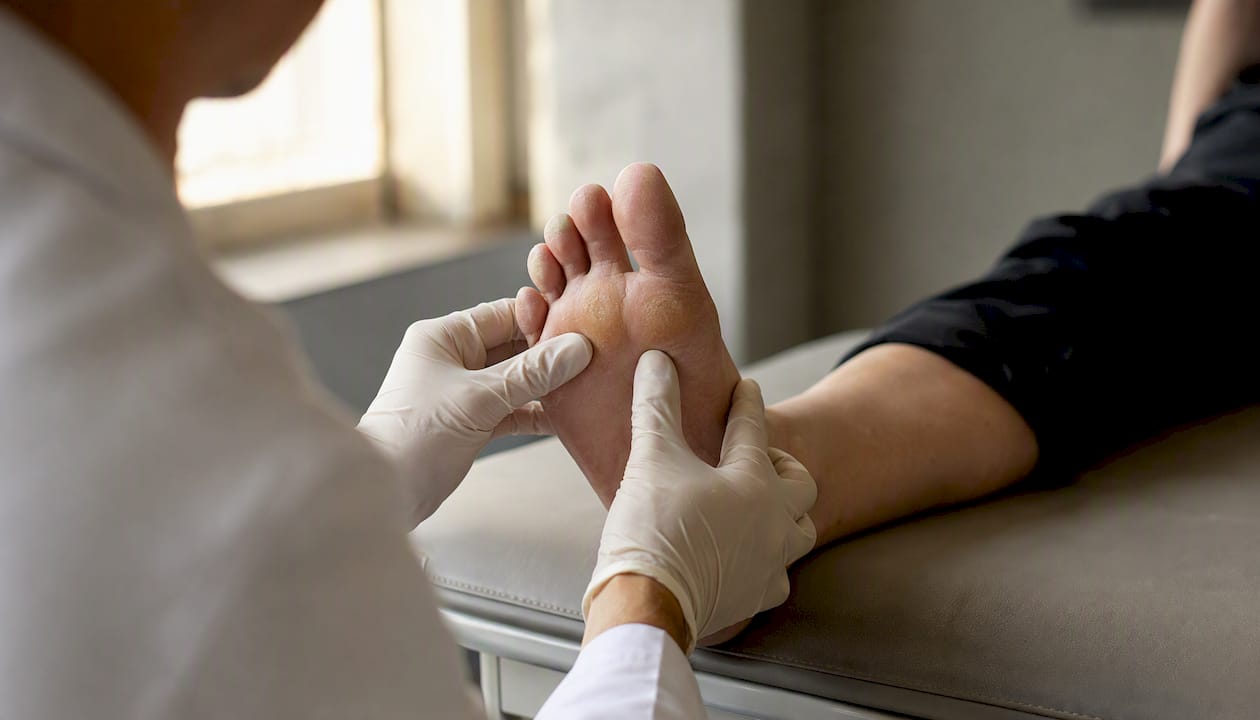
Objective testing: Gait observation and, where available, force-plate or in-shoe pressure mapping pinpoint which metatarsal head is overloaded and during which phase of gait. Imaging is ordered when a stress fracture, Freiberg’s osteonecrosis, or inflammatory joint disease is suspected, not routinely.
To prepare for your visit, review what to expect at your first appointment and bring the following:
Seek urgent evaluation if you have acute severe swelling after a specific incident, progressive numbness, or pain that is constant at rest, as these patterns suggest pathology beyond mechanical metatarsalgia.
Metatarsalgia is best understood as a load-management problem: identify the overloaded metatarsal head, determine the driver, and match the orthotic design accordingly. Clinical guidance identifies several common modifications, each with a specific indication.
| Modification | Primary indication | Expected effect on plantar pressure | Patient tolerance notes |
|---|---|---|---|
| Metatarsal dome (pad) | Central met head overload (2nd–4th) | Redistributes load proximal to met heads | Well tolerated; placement is critical |
| Forefoot cut-out | Focal lesion under single met head | Reduces peak pressure at that site | May reduce shoe fit in narrow toe boxes |
| Anterior shell drop-off | General forefoot offloading | Transfers load to proximal metatarsal shafts | Requires full-length shoe for best effect |
| First-ray cut-out | Hallux rigidus or first-ray overload | Reduces dorsiflexion resistance at first MPJ | Confirm first-ray mobility before applying |
| Heel lift (small) | Equinus/tight Achilles | Reduces forefoot compensation during gait | Pair with Achilles stretching program |
| Increased shell stiffness | Pes cavus, high-arch mechanics | Transfers load to arch and heel | May require longer break-in period |
Pro Tip: Pad placement is not intuitive. A metatarsal dome placed distal to the metatarsal heads, rather than proximal, can actually increase pressure and worsen pain. Always confirm placement with your clinician before leaving the fitting appointment.
Shell material matters too. A rigid polypropylene shell transfers load more effectively than a semi-flexible EVA shell, but patients with fat-pad atrophy often need a softer forefoot topcover added to the device for cushioning. Your clinician balances pressure redistribution against comfort based on your exam findings.
Orthotics manage load; rehab addresses the movement patterns that created the overload. Conservative care guidelines recommend combining orthotic devices with stretching, strengthening, and footwear modification for the best outcomes.
Home exercises (perform daily):
Clinic-based therapies to consider:
Shockwave therapy is used when chronic plantar tissue thickening or periosteal irritation is present and has not responded to exercise alone. Soft-tissue techniques such as Active Release Technique (ART) and Graston address restricted plantar fascia, intrinsic muscle tightness, and scar tissue that alter forefoot mechanics. Laser therapy may be added when local tissue inflammation is a primary component. Callus debridement by a clinician directly reduces peak pressure at the skin surface between orthotic adjustments.
Footwear is as important as the device itself. Your clinician will typically recommend shoes with a deep toe box, a low heel-to-toe drop, and a rocker sole profile. A rocker sole reduces forefoot loading during push-off, complementing the orthotic’s pressure redistribution.
Corticosteroid injections are occasionally used short-term to reduce acute inflammation, but they carry risks of fat-pad atrophy with repeated use. Your clinician will weigh that trade-off explicitly before recommending one.
The typical workflow runs: assessment → foot casting or 3D scanning → lab fabrication (usually 2–4 weeks) → initial fitting visit → follow-up at 2–6 weeks for adjustments.
At the fitting visit, your clinician checks shell contact, pad placement, and overall comfort in your shoes. Most patients are advised to begin with 2–4 hours of wear on day one and increase by one to two hours per day over 7–14 days. Rushing the break-in period is the most common reason for early discomfort.
Expect noticeable reduction in focal pressure and callus-related pain within the first few weeks of consistent wear. The meta-analysis cited earlier documented a mean plantar pressure reduction of -37.54 under the central metatarsal heads with bespoke orthotic treatment, and many patients report functional improvement within that same window. Full symptom resolution, particularly when combined with rehab, typically takes 8–12 weeks.
A successful trial means reduced pain with daily activities, improved tolerance for your trigger activities, and no new pressure points. If pain persists or migrates after 6–8 weeks of consistent wear, your clinician should reassess the design, check for footwear incompatibility, or reconsider the diagnosis.
Cost components at a U.S. clinic typically include the initial assessment visit, any casting or scanning fee, the orthotic fabrication fee, and follow-up adjustment visits. Fabrication fees vary by device type and lab.
Insurance coverage in the U.S. depends on your plan. Many commercial plans cover custom orthotics when medical necessity is documented, meaning your clinician must provide a diagnosis code, clinical notes, and a letter of medical necessity. Medicare covers therapeutic shoes and inserts under specific diabetic foot conditions; coverage for mechanical metatarsalgia varies by plan. Always confirm prior-authorization requirements before your appointment.
Before your visit, ask the front desk:
Bringing your insurance card and any referral documentation on your first visit speeds the process considerably.
Surgery is appropriate only after an appropriately dosed conservative trial has failed. That typically means persistent, disabling forefoot pain after several months of clinician-supervised orthotic use, targeted rehab, and footwear modification.
Seek prompt reassessment or referral if you notice:
Common surgical approaches include the Weil osteotomy (shortening and plantarflexion of a metatarsal), the DMMO (distal metatarsal minimally invasive osteotomy), and first-ray procedures for hallux-driven metatarsalgia. Each carries a recovery period of several weeks in a surgical shoe, with full return to activity typically taking 3–6 months.
Accurate diagnosis of the underlying driver, whether propulsive metatarsalgia from first-ray dysfunction, static overload from a long second metatarsal, or equinus-driven forefoot compensation, is critical before any surgical planning. Operating on the wrong driver produces poor outcomes regardless of technique.
The quality of your orthotic outcome depends heavily on the clinician’s assessment process, not just the device itself. When evaluating a clinic, look for:
Questions worth asking when you book:
For a broader look at what custom foot orthotics involve from a design and mechanics standpoint, partner clinic resources can supplement what your clinician explains at the visit.
Shephardhealth offers clinician-assessed custom foot orthotics integrated with gait and pressure assessment, shockwave therapy, laser therapy, and ART/Graston soft-tissue techniques. That combination means your orthotic design is informed by objective load data, and the complementary therapies are available in the same clinic if your recovery requires them.
Pro Tip: Ask specifically whether the clinic tracks your plantar pressure before and after orthotic fitting. Clinics that document outcomes, not just comfort scores, are better positioned to refine your device if the first version needs adjustment.
Most patients who come in with ball-of-foot pain have already tried cushioning products with limited success. The reason is almost always the same: the device was chosen before the driver was identified. Metatarsalgia is not a single condition. It is a symptom with several distinct mechanical causes, and the orthotic modification that helps one cause can actively worsen another. A heel lift for equinus-driven forefoot overload is the right call; that same lift in a patient with a long second metatarsal and no equinus adds nothing.
The evidence supports custom orthoses clearly, but the evidence also shows that the design details matter. Shell thickness, pad placement, and anterior edge geometry each change how load is transferred. Getting those details right requires a clinician who measures your pressure distribution, not one who estimates it. Patients who invest in a proper assessment and follow the break-in and rehab protocol consistently do better than those who treat the orthotic as a passive insert. The device is a tool; the clinical plan is what makes it work.
Persistent ball-of-foot pain deserves a clinical answer, not another trial-and-error product. At Shephardhealth, your first orthotic assessment includes a full physical exam, gait observation, and pressure analysis so your device is built around your actual load pattern, not a generic template. Shockwave therapy, laser therapy, ART, and Graston are available in the same clinic, meaning your treatment plan can expand without sending you elsewhere.

Direct insurance billing is available, and same-day appointments are offered for patients who need prompt evaluation. To get started, book your first visit and bring your most-worn shoes and any prior imaging. Your clinician will assess the driver of your pain, design a device matched to your mechanics, and set a clear follow-up schedule so you know exactly what to expect and when.
The claims in this article draw on the following clinical sources. Key quantitative findings are summarized below.
| Finding | Value | Source |
|---|---|---|
| Bespoke orthotic treatment: mean plantar pressure reduction under central met heads | MD -37.54, p=0.009 | OrthoArchives meta-analysis |
| Peak pressure reduction with optimized insole thickness | ~26.4% | Conservative Treatment for Primary Metatarsalgia |
| Custom orthoses vs. sham insoles in pes cavus | Significant reduction in forefoot pressure and improved pain/function | PROLAB Orthotics newsletter |
Annotated references:
This article provides general clinical information, not personalized medical advice. Confirm your diagnosis and treatment plan with a qualified clinician before beginning any orthotic or rehabilitation program.
Same Day Appointments Available
We Direct Bill!